3D Gallery
Look Closer: A Microscopy Exhibition
Enter gallery
Look Closer: A Microscopy Exhibition is a 3D virtual gallery on MyGallery3D, a walkable online exhibition of 13 works. Step inside and explore it in your browser: no app, no headset.
This 3D virtual museum of microscopy is one you walk through in your browser, so take your time at the glass.
Antonie van Leeuwenhoek was a draper in Delft who wanted to see the quality of his thread better than the lenses of his time allowed. He made more than 500 lenses, kept his method secret, and became the first person to see bacteria, spermatozoa and red blood cells. Nine of his microscopes survive, magnifying up to 275 times. It took about 150 years of optical development before the compound microscope could match them.
Living cells are nearly invisible under a bright field microscope, because their internal structures are colorless and transparent. The usual fix is to stain them, which often means killing and fixing the sample, and the dye can introduce artifacts, structures that are not really there. Frits Zernike's phase contrast, which won the Nobel Prize in 1953, images transparent living cells without any of that.
An electron's wavelength can be more than 100,000 times smaller than that of visible light, so a beam of electrons reaches what light cannot. Max Knoll and Ernst Ruska built the first electron microscope in 1931, and by 1933 their instrument beat the best optical one. Today the resolution is about 0.1 nm, and corrected lenses reach magnifications of more than 50 million times.
Visible light will not resolve anything smaller than about 200 nanometers. Push the magnification past roughly 1500x and the image gets bigger without getting truer: no new detail appears. That is diffraction, not bad glass. Every optical microscope ever built runs into the same wall, which is why the next step was to stop using light.
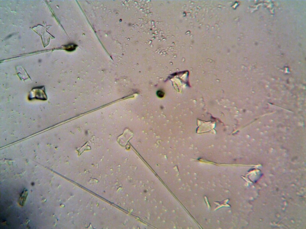
An assortment of particles captured at magnification. Each one presents its own distinct shape and surface texture, waiting to be examined more closely.
Photograph by turek, via Pexels.
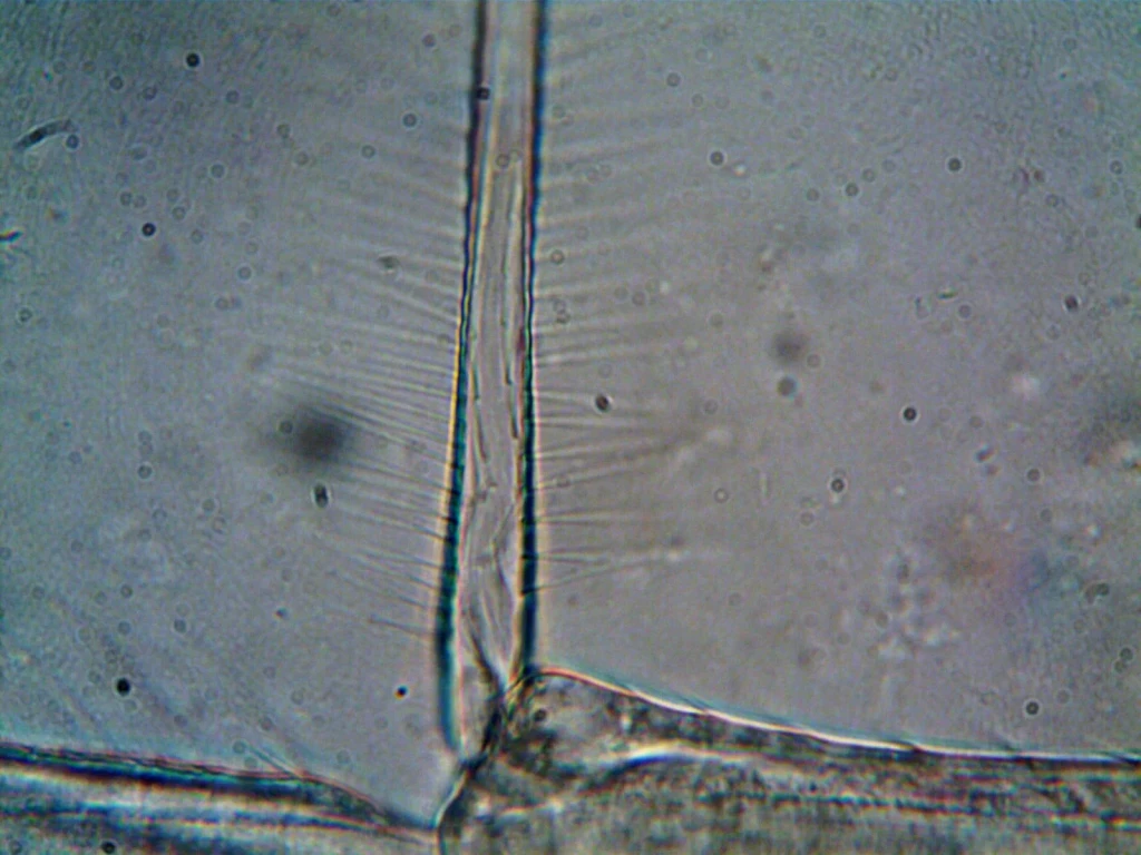
High magnification unveils intricate details of microorganisms. The exhibition's theme comes to life: there is always more to see.
Photograph by turek, via Pexels.
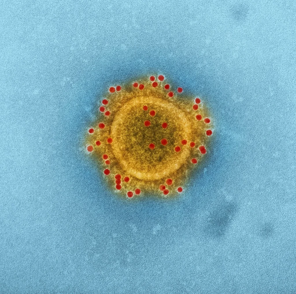
A high-resolution electron microscope image revealing the structure of a coronavirus particle. Magnification makes the invisible suddenly visible.
Photograph by CDC, via Pexels.
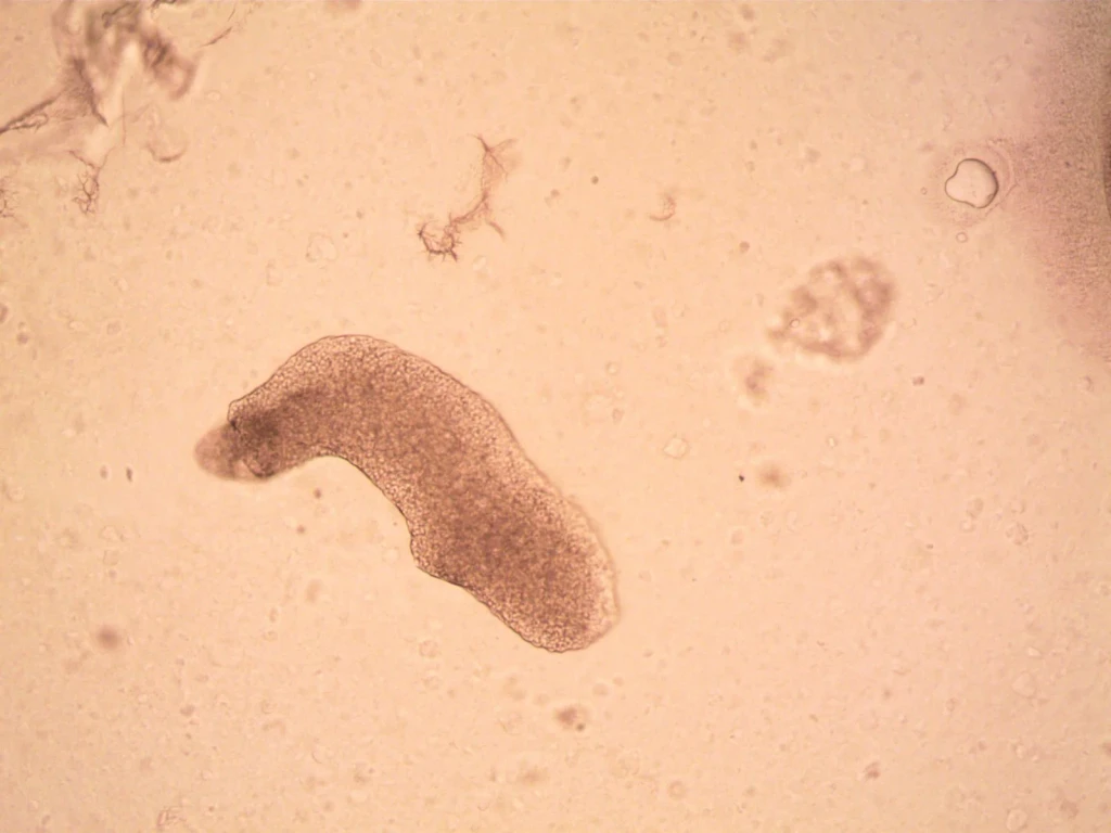
A microscopic animal in remarkable detail. The tardigrade's unique features become visible only when we look closer at life at this scale.
Photograph by turek, via Pexels.
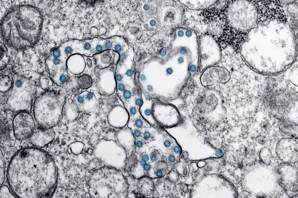
Gray spheres and blue elements arranged with flowing lines. An imagined view of the microscopic world, inviting us to consider how we visualize things too small to see.
Photograph by CDC, via Pexels.
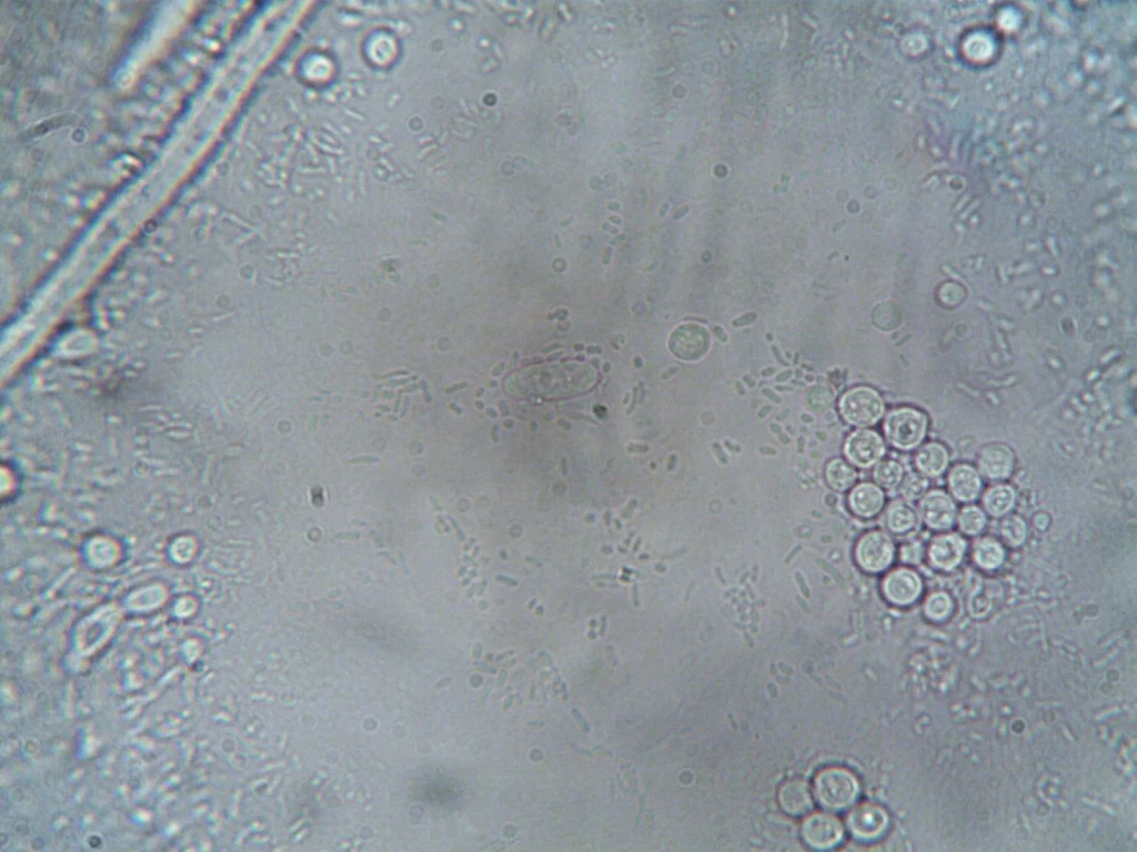
Various cellular structures and organisms revealed through magnification. Each detail tells a story about the complexity of microscopic life.
Photograph by turek, via Pexels.
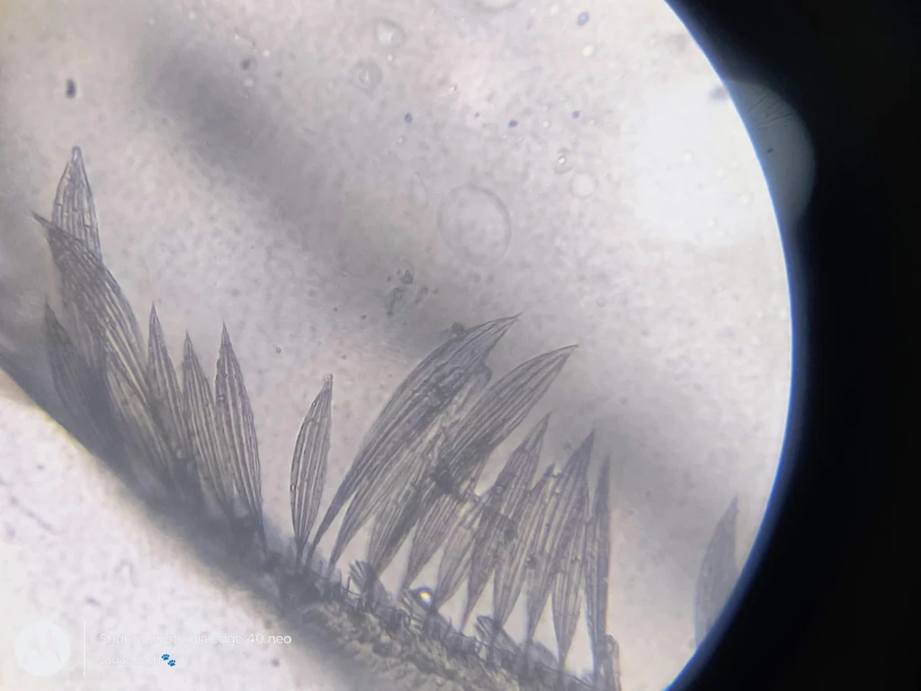
The feathery patterns of plant structure under the microscope. Nature's engineering becomes visible when we look closer at botanical design.
Photograph by Mahendra Jagadeesh, via Pexels.
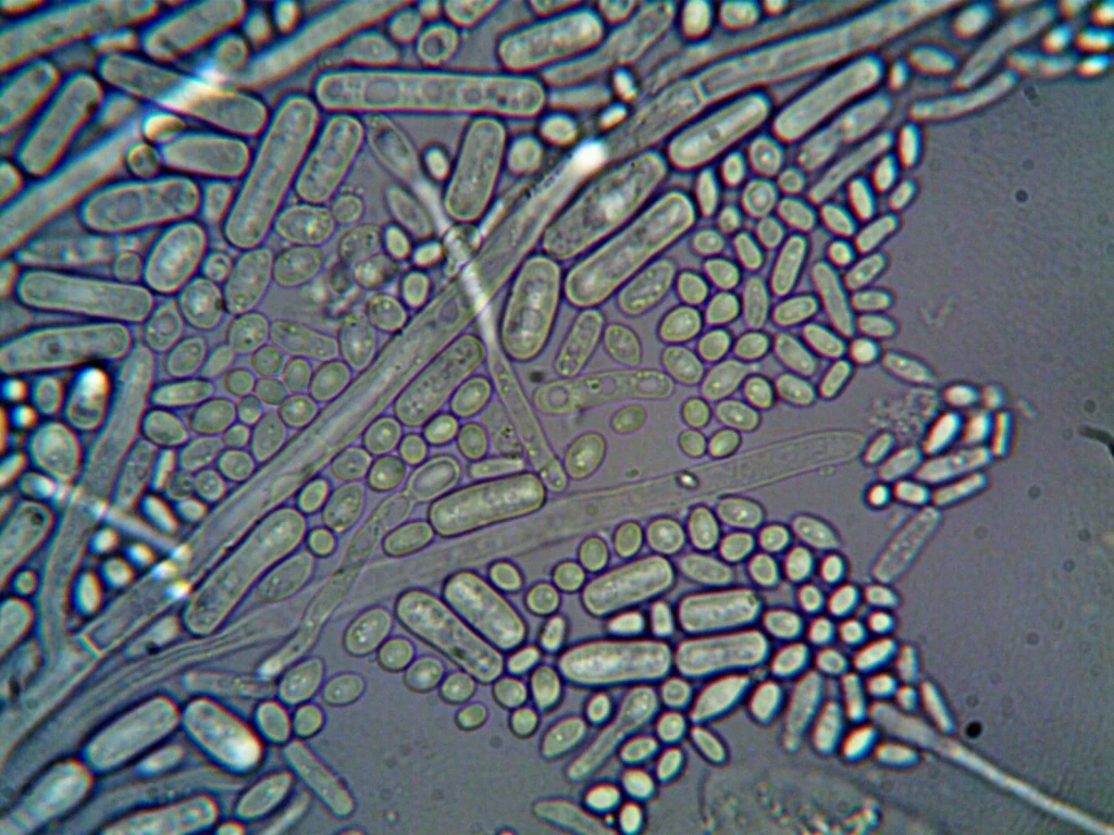
Cellular structures and organisms at high magnification. This image opens a window into a living world invisible to the naked eye.
Photograph by turek, via Pexels.
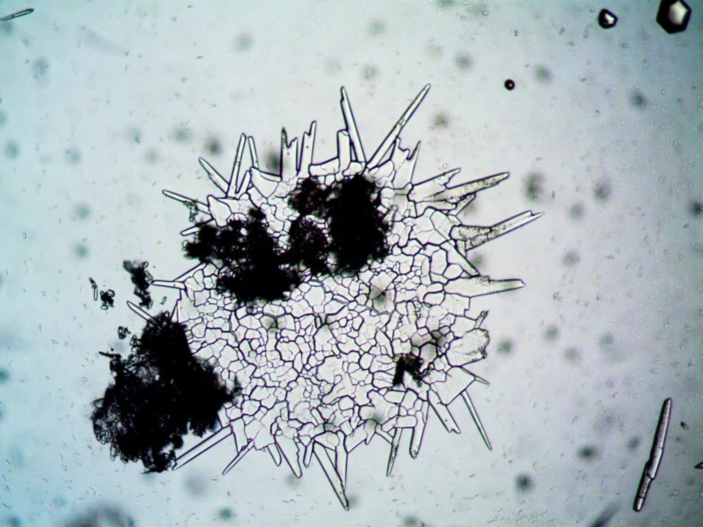
A cellular landscape of fungal spores and hyphae. This view reveals the intricate structures that fungi use to grow and spread through their environments.
Photograph by turek, via Pexels.
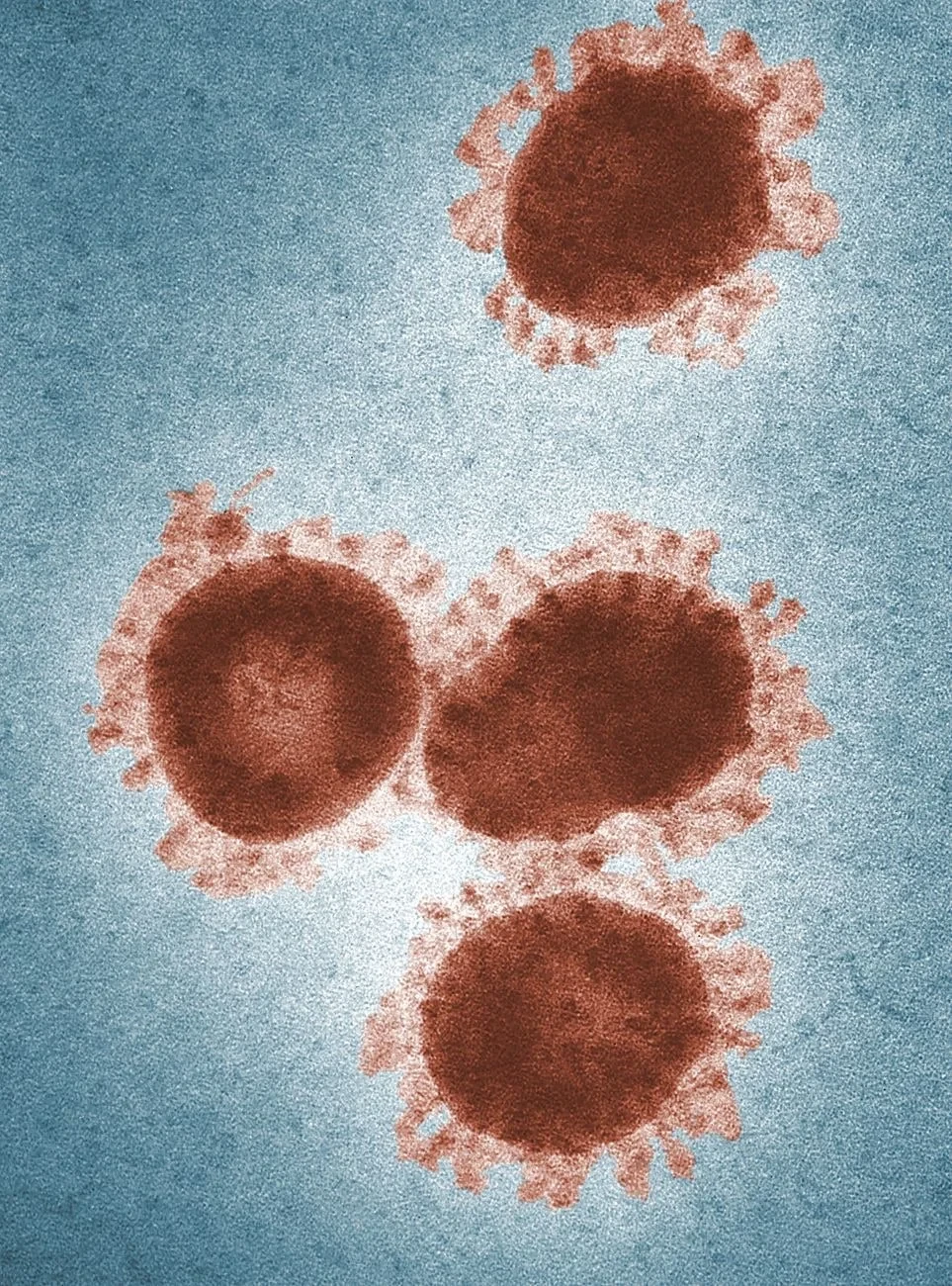
Color-enhanced electron microscopy shows coronavirus particles with their distinctive spike proteins clearly visible. Technology allows us to see what shapes our world.
Photograph by CDC, via Pexels.
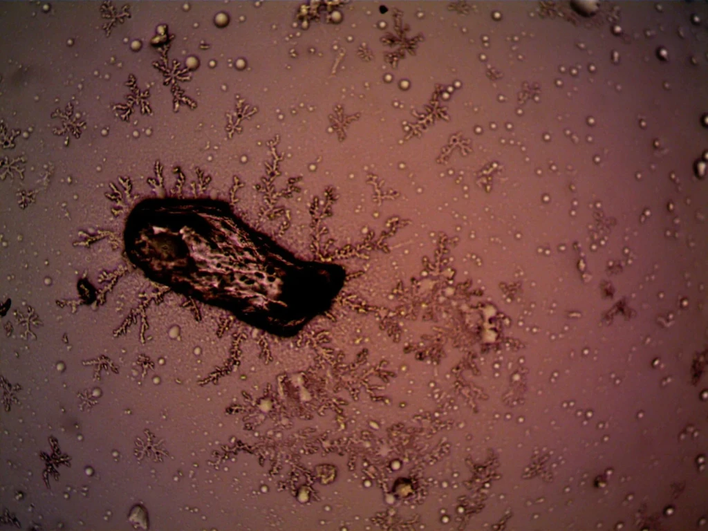
Bacteria and crystal formations create intricate patterns under magnification. Nature's complexity emerges at every scale.
Photograph by turek, via Pexels.
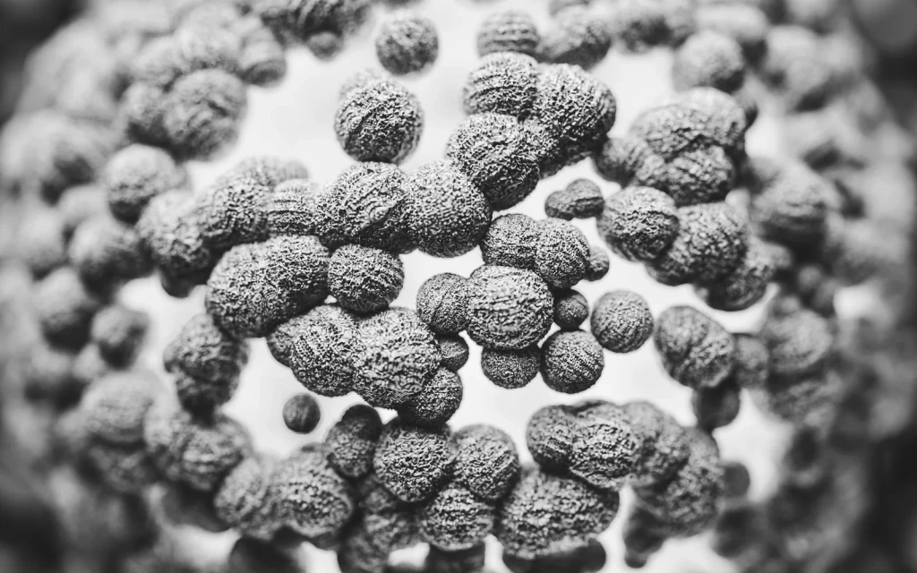
Black and white macro photography reveals the textured details of antibodies. Looking closer shows the elegant machinery of biological defense.
Photograph by Marek Piwnicki, via Pexels.
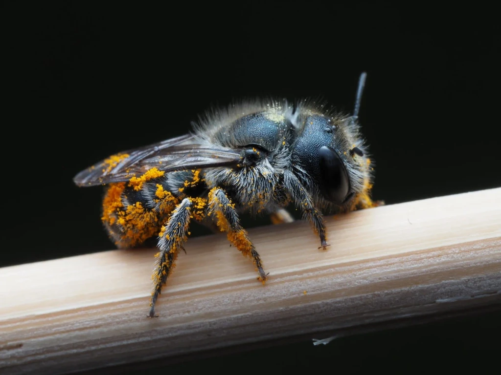
A bee becomes a study in texture and purpose. Magnified, its body reveals the intricate details of how life collects what it needs.
Photograph by Ali Goode, via Pexels.
